
Animal-Free Safety Assessment (AFSA)


Home_page_hero
Home_page_hero
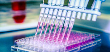
Imagine a world without animal testing… where the safety of chemicals, cosmetics, therapeutics and other products is assured using modern, human biology-based predictive tools. Making this world a reality is the goal of the AFSA Collaboration.
Imagine a world without animal testing… where the safety of chemicals, cosmetics, therapeutics and other products is assured using modern, human biology-based predictive tools. Making this world a reality is the goal of the AFSA Collaboration.
READ MORE
“I predict that 10 years from now, safety testing for newly developed drugs…will be largely carried out using human biochips…This approach…will mostly replace animal testing for drug toxicity and environmental sensing, giving results that are more accurate, at lower cost and with higher throughput.”
Francis Collins, MD, PhDDirector, U.S. National Institutes of Health 2016 READ MORE
The Animal-Free Safety Assessment (AFSA) Collaboration is a unique multi-sector partnership between non-profit, corporate, and philanthropic leaders working to advance acceptance and use of animal-free safety science as a gold standard across regulatory frameworks worldwide. Meet our members:
The Animal-Free Safety Assessment (AFSA) Collaboration brings together corporate and non-profit leaders who share the goal of accelerating a modern, species-relevant approach to safety assessment globally to better protect people and our planet, and hasten the replacement of animal testing. Meet our members:

Canada to phase out chemical toxicity testing on animals
The Canadian Government has passed historic amendments to the Canadian Environmental Protection Act, committing to developing a roadmap to prioritize animal-free testing methods and phase out chemical toxicity testing on animals by 2035.

Publication: Last resort requirement under REACH
Europe’s failure to uphold the legal requirement of chemical testing on animals as a “last resort” exposed in new publication.
South Korea to include recombinant Factor C in the pharmacopoeia
The synthetic alternative for the Bacterial Endotoxin Test will be added in the Korean Pharmacopoeia. Another country is recognizing the sustainable choice to move away from horseshoe crabs derived reagent.

REACHing for solutions
Watch our webinar and read our peer-reviewed paper outlining our recommendations to revise EU chemicals regulation and modernise safety assessment.

Biologicals publication: Event series on animal testing alternative for vet vaccines
In collaboration with IABS, and veterinary industry association, HelathforAnimals, we hosted an event series on the implementation and regulatory acceptance of non-animal testing for veterinary vaccines’ batch release testing.
Latest Events
-
The last resort requirement under REACH: From principle to practice
28 Mar 2024 -
Transition to non-animal based vaccine batch release testing. Policy and regulations theoretical aspects and case studies.
27 Mar 2024 -
12th World Congress on Alternatives and Animal Use in the Life Sciences
27 Aug 2023 -
Monocyte Activation Test the experience of the Italian Institute of Health
25 May 2023 -
Toward implementation plans for replacement of animal testing for human vaccines
19 Jan 2023 -
How to resolve inconclusive predictions from defined approaches for skin sensitisation in OECD Guideline No. 497
8 Dec 2022
REACHing for solutions: Essential revisions to the EU chemicals regulation to modernise safety assessment
Pereira M, Macmillan DS, Willett C, Seidle T.
Paving the way for application of next generation risk assessment to safety decision-making for cosmetic ingredients
Dent MP, Vaillancourt E, Thomas RS, Carmichael PL, Ouedraogo G, Kojima H, Barroso J, Ansell J, Barton-Maclaren TS, Bennekou SH, Boekelheide K, Ezendam J, Field J, Fitzpatrick S, Hatao M, Kreiling R, Lorencini M, Mahony C, Montemayor B, Mazaro-Costa R, Oliveira J, Rogiers V, Smegal D, Taalman R, Tokura Y, Verma R, Willett C, C. Yang
Systematic Review to Compare Chemical Hazard Predictions of the Zebrafish Embryotoxicity Test With Mammalian Prenatal Developmental Toxicity
Hoffmann S, Marigliani B, Akgün-Ölmez SG, Ireland D, Cruz R, Busquet F, Flick B, Lalu M, Ghandakly EC, de Vries RBM, Witters H, Wright RA, Ölmez M, Willett C, Hartung T, Stephens ML, Tsaioun KA